


Mastigopsis hjorti
Michael Vecchione and Richard E. Young

Introduction
Mastigopsis hjorti was originally described from five badly damaged specimens from the North Atlantic, then redescribed by Rancurel (1973) from three squid from the Gulf of Guinea. The species is distinctive and widely distributed but uncertainty exists on the taxonomic status of populations in other oceans.
Brief diagnosis:
A Mastigopsis ...
- with two photophores on each eyeball.
- with very large fins (length ca 90% of ML).
Characteristics
- Arms
- Arm III equals arm I in length (needs confirmation).
- Arm III equals arm I in length (needs confirmation).
- Tentacles
- Club suckers about equal sized except near tip.
- Club suckers about equal sized except near tip.
- Head
- Beaks. Description of the beaks can be found here in 2D.
- Beaks. Descriptions can be found here in 3D: Lower beak; upper beak.
- Funnel pocket absent.
- Funnel
- Funnel locking-apparatus with oval, slightly curved depression, posterolateral sides protude; without antitragus or clear tragus. Depression undercuts posterior margin.
- Fins
- Fins large; length about 90% of ML.
- Tubercules
- Large tubercules cover mantle, head, funnel and aboral surface of arms in subadults (tubercules are often lost during capture).
- Fins
- Fins large, nearly the full length of the mantle. (see title photograph).
- Photophores
- Two large circular photophores on ventral surface of eyeball; no other photophores present.
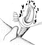
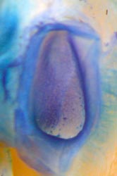
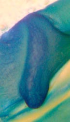
Figure. Frontal views of the funnel/mantle locking-apparatus of Mp. hjorti. Left - Funnel component (uppermost), mantle component (lowermost). Drawing from Rancurel (1973). Left two photographs - Funnel component (left), mantle component (right), equatorial Pacific. Right two photographs - Funnel component (left), mantle component (right), 73 mm ML, eastern North Atlantic, 17°24'N, 22°57'W, NMNH 815489. Tissue stained with methylene blue stain. Photographs by R. Young.
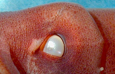
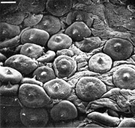
Figure. Left - Lateral view of head of Mp. hjorti showing tubercules and olfactory organ, 48 mm ML, western North Atlantic. Also visible are two lines on the head of the lateral-line analogue of cephalopods. Photograph by R. Young. Right - Scanning electron micrograph of mantle tubercles of Mp. hjorti, 93 mm ML, South Africa at 80°S, 05°E. Scale 0.1 mm. Photograph from Roper and Lu (1990).
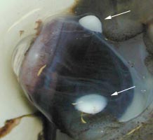
Figure. Ventral view of damaged eye of a fresh Mp. hjorti, western North Atlantic. Arrows point to photophores. Photograph by M. Vecchione.
Comments
More information on the description of Mp. hjorti can be found here.
Mp. hjorti bears resemblance to Idioteuthis cordiformis in the presence of large fins, skin tubercules, lack of a pocket between the bridles and the large trabeculate protective membranes on the tentacular clubs but differs in the presence of ocular photophores among other features.
Molecular Characteristics
Limited molecular data is available for this species (see Mastigoteuthidae: Discussion of Phylogenetic Relationships).Life History
Vecchione, et al. (2001) described a 6 mm ML paralarva which they assummed belonged to Mp. hjorti on the basis of a single large photophore on each eyeball. They described the paralarva as follows:
Mantle narrow, inserts on anterior end of fins. Fins ca. 25% of ML (excluding tail). Long, spike-like tail, nearly 3 times fin length. Skin mostly missing but fragments with scattered tubercules present. One light organ on ventral surface of each eye. Arm formula: II>I>IV>>III (arms III are minute buds). Tentacles long, thick, about 4 times length of arms II. Clubs with about 54 small suckers in 2 series proximally grading to 6 along "manus." Suckers end abruptly; tip with sucker anlagen.
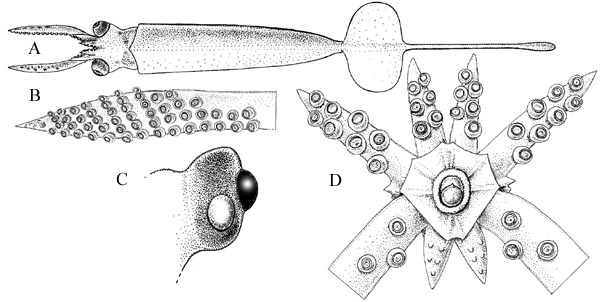
Figure. Paralarva of Mp. hjorti. A - dorsal view of paralarva, 6.0 mm ML, USNM 730521. B - oral view of tentacular club, same specimen. C - ventral view of eye with ocular light organ, same specimen. D - oral view of brachial crown, same specimen; note small arms III. Drawings from Vecchione, et al. (2001).
Distribution
Type locality: North Atlantic at 36°N, 40°W; 32°N, 33°W; 36°05'N, 43°58'W. The species is also known from the central Pacific (pers. obs.), off South Africa (Roper and Lu, 1990) and the Indian Ocean (Nesis, 1987).
References
Chun, C. 1913. Cephalopoda. Report on the Scientific Results of the "Michael Sars" North Atlantic Deep-sea Expedition 1910, 3(1). Reprinted by Bergen Museum, 1933, 21 pages.
Nesis, K. N. 1982/87. Abridged key to the cephalopod mollusks of the world's ocean. 385+ii pp. Light and Food Industry Publishing House, Moscow. (In Russian.). Translated into English by B. S. Levitov, ed. by L. A. Burgess (1987), Cephalopods of the world. T. F. H. Publications, Neptune City, NJ, 351pp.
Rancurel, P. 1973. Mastigoteuthis hjorti Chun 1913 description de trois échantillons proventant du Golfe de Guinée. Cah. O.R.S.T.O.M., ser. Océanogr., 11: 27-32.
Roper, C.F.E. and C.C. Lu 1990. Comparative morphology and function of dermal structures in oceanic squids (Cephalopoda). Smithson. Contr. Zool., No. 493: 1-40.
Vecchione, M., C. F. E. Roper, M. J. Sweeney and C. C. Lu. 2001. Distribution, relative abundance and developmental morphology of paralarval cephalopods in the western north Atlantic Ocean. NOAA Technical Report NMFS 152: 1-58.
Title Illustrations
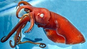
| Location | Western North Atlantic, 24°N, 82°W |
|---|---|
| Comments | Captured with a slurp gun from the Johnson Sea Link II submersible. |
| Specimen Condition | Preserved |
| View | Side |
| Size | 48 mm ML |
| Image Use |
 This media file is licensed under the Creative Commons Attribution-NonCommercial License - Version 3.0. This media file is licensed under the Creative Commons Attribution-NonCommercial License - Version 3.0.
|
| Copyright |
© 2004

|
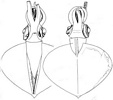
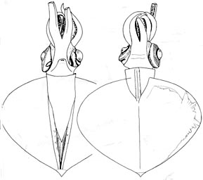
| Scientific Name | Mastigoteuthis hjorti |
|---|---|
| Location | Gulf of Guinea |
| Comments | Ventral and dorsal views. |
| Reference | Rancurel, P. 1973. Mastigoteuthis hjorti Chun 1913 description de trois échantillons proventant du Golfe de Guinée. Cah. O.R.S.T.O.M., ser. Océanogr., 11: 27-32. |
| Size | 44 mm ML |
About This Page
National Museum of Natural History, Washington, D. C. , USA
University of Hawaii, Honolulu, HI, USA
Page copyright © 2019 and
 Page: Tree of Life
Mastigopsis hjorti .
Authored by
Michael Vecchione and Richard E. Young.
The TEXT of this page is licensed under the
Creative Commons Attribution-NonCommercial License - Version 3.0. Note that images and other media
featured on this page are each governed by their own license, and they may or may not be available
for reuse. Click on an image or a media link to access the media data window, which provides the
relevant licensing information. For the general terms and conditions of ToL material reuse and
redistribution, please see the Tree of Life Copyright
Policies.
Page: Tree of Life
Mastigopsis hjorti .
Authored by
Michael Vecchione and Richard E. Young.
The TEXT of this page is licensed under the
Creative Commons Attribution-NonCommercial License - Version 3.0. Note that images and other media
featured on this page are each governed by their own license, and they may or may not be available
for reuse. Click on an image or a media link to access the media data window, which provides the
relevant licensing information. For the general terms and conditions of ToL material reuse and
redistribution, please see the Tree of Life Copyright
Policies.
- First online 18 July 2004
- Content changed 31 October 2018
Citing this page:
Vecchione, Michael and Richard E. Young. 2018. Mastigopsis hjorti . Version 31 October 2018. http://tolweb.org/Mastigopsis_hjorti/19517/2018.10.31 in The Tree of Life Web Project, http://tolweb.org/


