
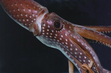
Figure. Right-ventral view of the head of H. cerasina. Photograph by R. Young.
- Photophores (head)
- Compound photophores (Type 2a): Click on an image to view larger version & data in a new window
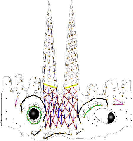
Figure. Arrangement of photophores on head and arms. Dorsal, lateral and ventral views combined in the illustration. The vertical line on either side of head in illustration represents the dorsal midline of the head. Photophore groups are connected by color-coded lines. Drawing by R. Young.
- Basal Arm IV Row with 3 photophores. Medial photophore separated by 2 photophores from anterior photophore of Midline Series.
- Midline Series with 2 photophores.
- Right Eyelid Series with 17 photophores .
- Left Eyelid Series with 6 photophores.
- 2° Right Eyelid Series with 8 photophores.
- 2° Left Eyelid Series with 7 photophores.
- Ventral Matrix with 42 photophores; 11 longitudinal (thin blue) series; attachment to 2° Eyelid Series at 2° eyelid photophores no. 2 on either side.
- Ventral Matrix photophores tend to align in transverse rows.
- Right basal series with 2 photophores.
- Left basal series with two very small photophores.
- Basal row smooth alignment with 7 photophores .
- Right Accessory Series with 2 photophores (see below).
- Left Accessory Series with 3 photophores (see below).
Simple photophores (round, black dots):
- Arrangement as illustrated.
- A few slightly enlarged, simple photophores on dorsal surface of head.
- Small, simple photophores not easy to recognize and may be variable.
- Compound photophores (Type 2a):
- Compound photophores (mantle)
- Ventral mantle with about 10 photophores in a diagonal row.
- Photophores in anterior half of ventral mantle approximately uniform in size.
Click on an image to view larger version & data in a new window
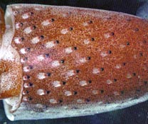
Figure. Ventral view of the mantle of H. cerasina. Left - Holotype. Drawing from Nesis, 1971. Right - Live squid, from Hawaiian waters. Photograph by R. Young.
- Compound photophores (arms)
- Arm tip without separated group of compound photophores (top right photograph, drawing at top of page).
- Arms IV with photophores in 3 series in proximal 2/3 (bottom right photograph).
- Arms III with two longitudinal series at bases (drawing at top of page).
- Arms II with two longitudinal series at bases (lateral two series barely separable) (drawing at top of page).
- Arms I with one longitudinal series at bases (drawing at top of page).
Click on an image to view larger version & data in a new window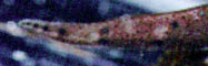
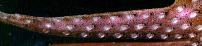
Figure. Ventral view of arm IV of H. cerasina. Top - Arm tip, preserved squid, 60 mm ML, Hawaii. Bottom - Most of arm IV, same live squid as above, Hawaii. Photograph by R. Young.
- Tentacle
- Club length 10-15% of ML.
- Locking apparatus with 8-10 suckers; formula: s, k, s, s, k, k, s, s, 2k, 2s, s, k, s, k, k, //, k, s, (k), s, (k). k=knob, s=sucker, //=border of club manus. Click on an image to view larger version & data in a new window
Figure. Oral view of tentacle of H. cerasina, holotype. Drawing from Nesis, 1971.
- Club suckers with 6 irregular series in central portion of club.
Click on an image to view larger version & data in a new window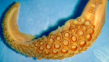
Figure. Oral view of club of H. cerasina, est. 100 mm ML, Hawaii, preserved. Photograph by R. Young.
- Suckers (club)
- Largest suckers of large specimens 4-5% of ML in diameter.
- Largest suckers with small, sharp, narrow teeth around entire border (photographs on the left; drawing on the right); teeth number 30-35 in small squid (34 mm ML) and 40-60 in large squid (48-52 mm ML).
Note that in the photograph the distal teeth are longer and more slender than the proximal teeth. The sucker on the left has about 42 teeth (ca. 100 mm ML).
Click on an image to view larger version & data in a new window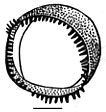
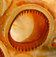
Figure. Oral view of suckers from tentacular club of H. cerasina. Left - inner ring of largest sucker, paratype, 34 mm ML. Drawing from Nesis, 1971. Right - Large sucker, mantle missing, estimated size 100 mm ML, Hawaii, preserved. Photograph by R. Young.
- Suckers (arm)
- Diameter of largest arm suckers about 2.5% of ML.
- Suckers of arms I-III with smooth inner rings (photograph to near right); suckers of arms IV with disorderly arranged, low, blunt teeth (photograph to far right).
Click on an image to view larger version & data in a new window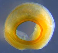
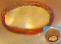
Figure. Oral view of large arm suckers of H. cerasina. Left - Sucker from arm III, 60 mm ML, Hawaii, preserved. Right - Sucker from arm IV, close-up of inner ring and insert of whole sucker, 60 mm ML, Hawaii, preserved. Photographs by R. Young.
- Arms and web
- Arm lengths 100-150% of ML; subequal with arms IV slightly smaller.
- Inner web 15% of longest arm; outer web absent.
- Occipital folds
- Two barely detectable folds on either side of head.
- Funnel organ
- Arms of funnel organ without ridges or lobes.
Click on an image to view larger version & data in a new window
Figure. Ventral view of funnel organ of H. cerasina, paratype, 52 mm ML. Drawing from Nesis, 1971.
- Fins
- Length 30-40% of ML.
- Width 50-68% of ML.
Comments
Except for the analysis of the head photophores and the photographs, this description is mostly from Nesis (1971).


