UPPER BEAK CHARACTERISTICS:
- Step: Present in Type 1 and Type 3 beaks, absent in Type 2 beaks. Located between Palatoshoulder Ridge (or occasionally Hyaline Matrix) and Jaw Edge in beak Types 1 and 3.
- Shoulder Blade: Thin; on Outer Part of Shoulder, without an Outer Cover.
- Shoulder Blade: Presumably composed of a single Component.
- Hyaline Matrix: Forms Middle Part of Shoulder.
- Palatoshoulder Ridge: Forms Inner Part of Shoulder except in Type 3 beaks where it is virtually absent.
- Pigmentation of posterior surface of Hyaline Matrix: Begins on posteromedial side of Hyaline Matrix with the LW-B Continuum appearing to "peal-off" of Lateral Wall.
- Yellow Line: Absent.
Bipartite Upper Beak: Structure
- EXTERNAL FEATURES
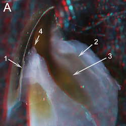
- The major parts. In moderately pigmented Bipartite beaks (i.e., beaks of intermediate age) we can recognize the same three parts (i.e., groups of very thin layers) of the Shoulder seen in Vampyoteuthis: (1) An outer (lateral) part which contains the pigmented Shoulder Blade; (2) a middle part composed of the thick region of unpigmented Hyaline Matrix; and (3) an inner part consisting of the pigmented Palatoshoulder Ridge of the Lateral Wall. The two pigmented parts give this beak type its name (Bipartite).
 Click on an image to view larger version & data in a new window
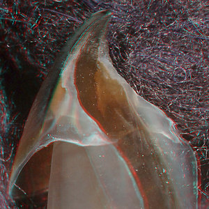
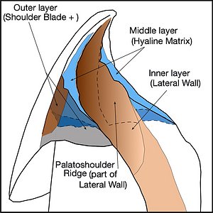
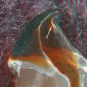
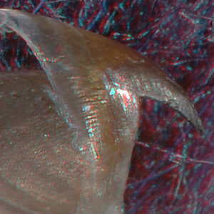
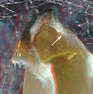
Figure. Oblique views of Magnoteuthis microlucens, immature female, 95 mm ML, 3.2 mm URL. Left - 3D photograph. Right - Drawing taken from the photograph. The dashed line that defines two sides of the Palatoshoulder Ridge indicates where the Ridge merges with the rest of the Lateral Wall posteriorly and with the Bridge dorsally. Images by R. Young.
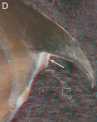
- The Step. Bipartite beaks lack a Step early in development but most develop one later. In some cases the Step may not develop until the beak is mostly pigmented. In relatively young beaks (i.e., prior to the development of a Step) the free, anterior edge of the Shoulder Blade is the most anterior component of the Shoulder. Typically a Step forms during ontogeny by regression (often erosion) of the free (= anterior) edge of the Shoulder Blade which (since the Shoulder Blade is fused to the Hood at the Jaw-edge Extension) extends the Jaw Edge posteriorly; a Step (i.e., a gap) is, thereby, formed between the Palatoshoulder Ridge (or in some cases the Hyaline Matrix) and the Jaw Edge. The size of the Step is accentuated by the continued growth of the Palatoshoulder Ridge. In the few taxa without a Step, the Shoulder Blade does not regress and the anterior edge of the Palatoshoulder Ridge never surpasses that of the Shoulder Blade.
- The Shoulder Blade. The pigmented Shoulder Blade is smoothly attached to the Jaw Edge Extension. The pigmentation usually fades posteriorly. The Shoulder Blade can be very narrow (dorsal-ventral direction) and sometimes difficult to detect, or it can occupy over half of the outer part of the Shoulder. The Shoulder Blade generally lacks any external covering.
- The Palatoshoulder Ridge. The Lateral Wall that forms the inner layer of the Shoulder often extends well anterior to the rest of the shoulder to form "free edges" that can extend onto the Palate. On the Palate, the Lateral Wall is an extension of the Median Pallet (i.e., the middle part of the pallet) and connects laterally to the Lateral Palate (i.e., the portion of the Bridge that lies on the Palate) which, in turn, connects with the Hood. The Palatoshoulder Ridge, therefore, is simply a continuation of the pigmented Lateral Wall. It is defined dorsally by its junction with the Bridge and posteriorly by its junction with the B-LW Continuum (LateralWall-Bridge Continuum) or, in poorly pigmented beaks, by the posterior termination of the Hyaline Matrix (see drawing above).
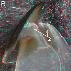
- The types of Bipartite Beaks. The structure of the Shoulder changes greatly during ontogeny. When the Shoulder Blade and the Lateral Wall first become pigmented, the Shoulder Blade is usually the more anterior part of the Shoulder preventing the presence of a Step. As the squid grows, the Shoulder Blade often recedes/erodes revealing a new surface of the Jaw Edge. Usually the Hyaline Matrix also recedes. In either case a Step is created between the Jaw Edge and either the Hyaline Matrix or the developing Palatoshoulder Ridge. The result is that the squid changes during ontogeny from having a beak without a Step to one with a Step (the latter change is a primay feature of a Bipartite Type 1 beak). In some species the Shoulder Blade doesn't recede and a Step never develops. The Shoulder Blade, therefore, remains the anterior-most structure of the Shoulder (a Bipartite Type 2 beak). In the Bipartite Type 3 beak the Palatoshoulder Ridge never develops. The developmental stage of the beak as seen in its degree of pigmentation, therefore, is important in identifying the type of Bipartite beak.
- Bipartite Type 1: Step present laterally between Palatoshoulder Ridge (or sometimes Hyaline Matrix) and Jaw Edge at some point during ontogeny.
- Biparatite Type 2: Step absent. Palatoshoulder Ridge present but not anterior to Shoulder Blade which generally precludes Step development.
- Bipartite Type 3: Step present. Palatoshoulder Ridge virtually absent, apparently appearing only with general pigmentation of Shoulder. See Bipartite UB Survey
Click on an image to view larger version & data in a new window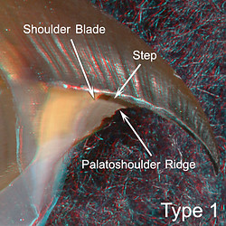
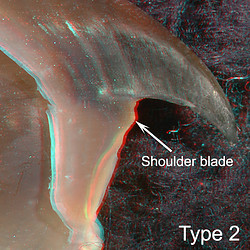
Figure. Bipartite beak types. Left - Gonatus berryi, 124 mm ML, ? mm URL. The regression of the Shoulder Blade has left the Palatoshoulder Ridge in a relatively more anterior position creating a Step (accentuated in the image by a shadow). Right - Berryteuthis magister, ? mm ML, 5.9 mm URL. Although the Shoulder Blade is unrecognizable due to the general pigmentation of the Outer Part of the Shoulder, no regression is apparent.
- The major parts. In moderately pigmented Bipartite beaks (i.e., beaks of intermediate age) we can recognize the same three parts (i.e., groups of very thin layers) of the Shoulder seen in Vampyoteuthis: (1) An outer (lateral) part which contains the pigmented Shoulder Blade; (2) a middle part composed of the thick region of unpigmented Hyaline Matrix; and (3) an inner part consisting of the pigmented Palatoshoulder Ridge of the Lateral Wall. The two pigmented parts give this beak type its name (Bipartite).
- INTERNAL STRUCTURE: Longitudinal cuts.
A longitudinal-cut through the Shoulder aids in understanding the shoulder structure (images below). The cut was made with a scalpel and photographed (in 3D) at two slightly different angles to aid the viewer in understanding the orientation in the succeeding photomicrograph. The latter, taken through a stereomicroscope (in 2D), shows the most detail.
The Shoulder Blade and the Lateral Wall have a very simple structure and we designate the former as a "single component" Shoulder Blade. The Hyaline Matrix is very thick compared to the thickness of the adjacent pigmented structures. This beak was on the verge of developing a Step.Click on an image to view larger version & data in a new window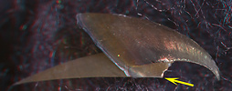
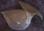
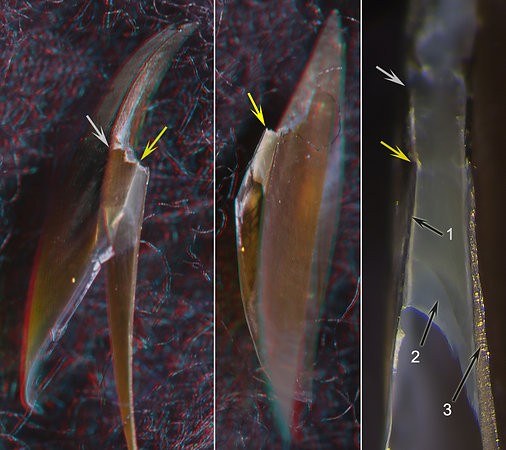
Figure. Magnoteuthis microlucens, immature male, 115 mm ML, 2.2 mm URL. TOP: Left - Side view showing where the longitudinal cut was made. Right - Side view of the whole beak showing the extent of pigmentation. BOTTOM: Cut beak with rostrum directed upward. Left - Oral-lateral view showing the Shoulder Blade. Middle - Oral-medial view showing the Palatoshoulder Ridge. Right - Photomicrograph (2D), oral view, taken perpendicularly to the cut surface. White arrows - Point to the Jaw Angle. Yellow arrows - Point to same spot on all beaks (anteriolateral end of the Shoulder Blade where cut was made). Arrow 1 - Shoulder Blade. Arrow 2 - Hyaline Matrix. Arrow 3 - Palatoshoulder Ridge.
- INTERNAL STRUCTURE: Cross-sectional cuts.
A cross-cut through the shoulder gives a very different view of the shoulder structure (figure below). The cut was made with a scalpel and photographed (in 3D) at two slightly different angles to aid in understanding the orientation of the following photomicrograph. The latter, taken through a stereomicroscope (in 2D), shows the most detail.
Click on an image to view larger version & data in a new window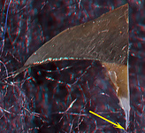
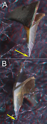
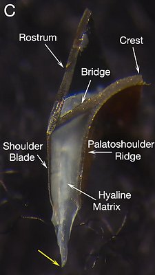
Figure. Magnoteuthis microlucens, immature male, 115 mm ML, 2.2 URL, showing cross-sectional cut through the shoulder. TOP - Side view of a beak fragment showing where the upper beak was cut. BOTTOM: A - Posterolateral view showing cut surface and Shoulder Blade. B - Posteromedial view showing cut surface and Palatoshoulder Ridge. C - Photomicrograph (2D) of cut region. Yellow arrows - indicate same spot on all images.
- INTERNAL STRUCTURE: Views of a beak without Hyaline Matrix
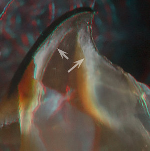
To better understand the structure the Bipartite beak, examine the images below of a Type 1 beak in which all hyaline material has been dissolved away leaving the pigmented "skeleton." The LW-B Continuum appears to "peal off" the edge of the Lateral Wall to become, dorsally, a continuation of the Bridge and posteriorly the cover of the posteromedial surface of the missing Hyaline Matrix. This cover is, however, incomplete at this stage as a gap still exists (Fig. C, E1). The internal limits of the Palatoshoulder Ridge (Figs. A, B, D) can be seen where the Lateral Wall intersects the LW-B Continuum and where the Ridge dorsally intersects the Bridge. In Type 2 beaks the role of the Palatoshoulder Ridge is reduced. The presence of a LW-B Continuum is a characteristic of all decapodiform beaks (absent in Vampyroteuthis) but varies considerably in different types of beaks.
Click on an image to view larger version & data in a new window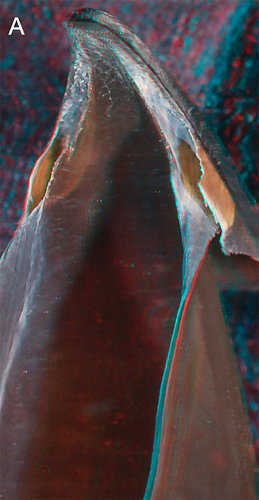
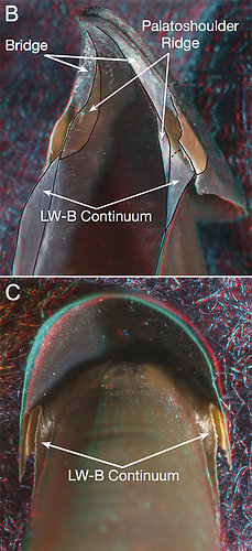
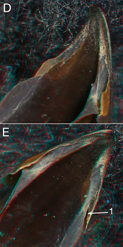
Figure. Different views of the upper beak of Magnoteuthis magna, 141 mm ML, 3.3 mm URL, with hyaline material dissolved away. A - Oblique view showing the basic features of the beak. B - Same picture with the basic parts outlined in black and labeled. C - Posterior view showing the LW-B Continuum nearly covering the posterior region of the middle shoulder. D, E - Two more views of the same beak showing how the LW-B Continuum merges with the Bridge. Arrow 1 (Fig. E) - The gap in the posterior pigment covering of the missing Hayline Matrix. In slightly older beaks this gap is closed.
- CHANGES WITH GROWTH: Magnoteuthis microlucens (mostly)
? sex, 26 mm ML, 0.32 mm URL
? sex, 60 mm ML, 1.6 mm URL Immature female, 95 mm ML, 3.2
mm URL
Mature male, 215 mm ML, 4.7 mm
URL
Side
view
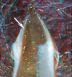
A - At 0.32 mm URL (very small), the beak has a well pigmented Shoulder Blade (arrow) whose anterior edge is the most anterior element in the Shoulder. B - At 1.6 mm URL, the Palatoshoulder Ridge (arrow) is very slightly anterior to the Shoulder Blade (the white material at the front of the Shoulder Blade is some Hyaline Matrix, the anterior fringe of the Ridge is just visible behind it. C - At 3.2 mm URL, the Palatoshoulder Ridge (arrow) is distinctly the most anterior structure of the Shoulder, creating a Step between it and the Jaw Edge. Note that the step is accentuated by a shadow. D - In the heavily pigmented mature male (4.7 mm URL) the original Shoulder Blade is reduced and lost within the fully pigmented outer layer of the Shoulder. The Palatoshoulder Ridge (arrow) is well anterior of the other Shoulder elements, forming a large Step.
- UNUSUAL BIPARTITE BEAKS: Grimalditeuthis bonplandi
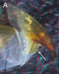
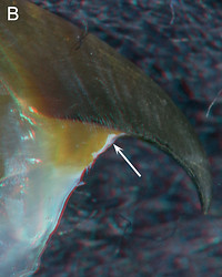
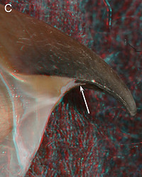
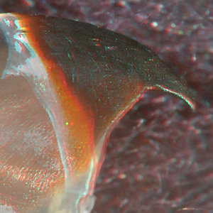
G. bonplandi has a very peculiar Bipartite beak. It has virtually lost its Shoulder Blade (A, 1) which, generally, is marginally detectable in only young beaks. The Palatoshoulder Ridge (B, 2) is medial to the Hyaline Matrix (as typical of Bipartite beaks) but its medial surface is covered by hyaline material; the Ridge emerges anteriorly (C, 2) from this cover in very large beaks. The Ridge, therefore is mostly composed of only the outer, pigmented layers of the Lateral Wall (see Internal Structure below). The anterior edge of the Ridge has an unusual shape in beaks of intermediate size (B, 2) and mostly is absent medial to the embayment of the Hyaline Matrix. At this size there is little indication of a Ridge on the Patale but is the larger beaks (137 mm ML, 3.0 URL and C above) a short Ridge is clearly present and also forms a medial base to the Hyaline Matrix embayment.
Grimalditeuthis bonplandi, 22 mm ML, 1.3 URL, oblique view
G. bonplandi, 120 mm ML (estimate), 1.8 mm URL. oblique view.
G. bonplandi, 155 mm ML, 2.6 mm URL, oblique view.
ARROWS (all images above and below): 1 - Shoulder Blade. 2 - Palatoshoulder Ridge. 3 - Shadow cast by opposite side of beak. 4 - Hyaline Matrix (in A & B the dark color is a lighting artifact). 5 - Hyaline material covering the medial surfaces of the Ridge and Palate. 6 - Lateral edge of cut and of Hyaline Matrix. 7- Hood. 8 - Bridge. 9 - Palate. Yellow arrows - Indicate to same spot on beaks in all D-images and, separately, all E-images.
- INTERNAL STRUCTURE: Grimalditeuthis beak - Longitudinal and cross-sectional cuts. Click on an image to view larger version & data in a new window
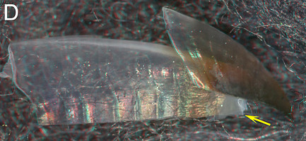
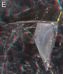
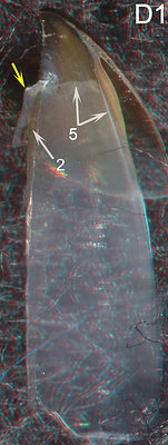
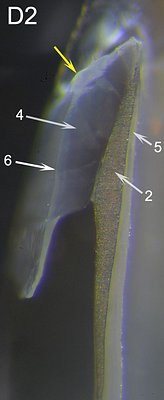
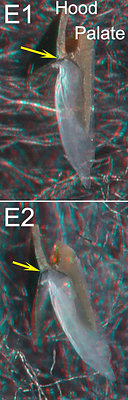
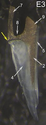
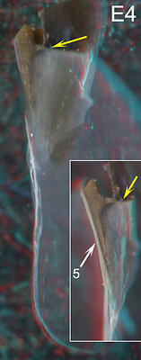
Figure. Grimalditeuthis bonplandi, ? mm ML, 1.8 mm UL. D-series: D - Side view of cut beak showing position of the longitudinal cut. D1 - Oral-medial view of cut beak. Note the abrupt anterior end of the hyaline material (5). D2- Photomicrograph (2D) of cut, oral view, taken perpendicular to the cut surface. Note that the Palatoshoulder Ridge (2) is a thickening of the pigmented outer layer of the Lateral Wall.
E-series: E - Side view of cut beak showing position of cross-sectional cut. Note the very different size of the embayment in D and E. E1 - Posterolateral view of anterior piece of cut beak. E2 - Posterolateral view with a dorsal tilt of anterior piece of cut beak. E3 - Photomicrograph (2D) of cut, oral view, taken perpendicular to cut surface of anterior piece of cut beak. Note the absence of a recognizable Shoulder Blade. E4 - Two slightly different anterolateral views of posterior piece of cut beak. The insert caught the light just right to show 5.
| sex ?, 26 mm ML, 0.32 mm URL | sex ?, 60 mm ML, 1.6 mm URL | Immature female, 95 mm ML, 3.2 mm URL | Mature male, 215 mm ML, 4.7 mm URL | |
| Oblique |  | 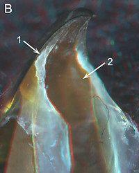 | 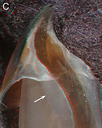 | 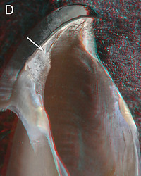 |
A - At 0.32 mm URL the Lateral Wall (arrows) is pigmented dorsal and posterior to the Shoulders but not on the Shoulders. The Palatoshoulder Ridge is not yet present. B - At 1.6 mm URL the beaks show an expanded pigmentation of the Lateral Wall with a well-developed Palatoshoulder Ridge (arrow 2) and a distinct Step (arrow 1). C - At 3.2 mm URL the beak has changed little except for the greater lateral pigmentation of the Lateral Wall (arrow). D - At 4.7 mm URL the Hyaline Matrix is sclerotized and shrunken. The white material on the surface of the Shoulder and the rostrum (inner and outer surfaces of this beak is of unknown origin and composition. However, it is a hard substance and should not be confused with the translucent and relatively soft Hyaline Matrix. With the Hyaline Matrix virtually gone, the Step (arrow), which extends posteriorly to the Jaw Angle, is much more apparent.
| sex ?, 26 mm ML, 0.32 mm URL | sex ?, 60 mm ML, 1.6 mm URL | M. hjorti, Immature female, 115 mm ML, 3.8 mm URL | Mature male, 215 mm ML, 4.7 mm URL | |
| Posterior | 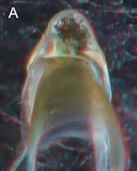 | 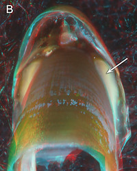 | 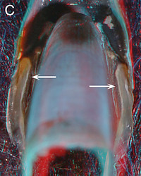 | 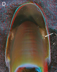 |
A - At 0.32 mm URL, the posterior view of the beak, the Shoulder is not very apparent. However, the extent of the dorsal and posterior pigmentation of the Lateral Wall is clear. B - At 1.6 mm URL, the distinct Hyaline Matrix (arrow) is covered dorsally by the pigmented Bridge and laterally it abuts the Shoulder blade. C - The 3.2 mm URL version of M. microlucens was virtually the same as that seen in B. We substituted a slightly more advanced version of M. hjorti which shows pigmentation beginning to cover the Hyaline Matrix arising primarily from its medial side as part of the LW-B Continuum (arrows). D - At 4.7 mm URL, the posterior surface of the Hyaline Matrix is fully pigmented.
Occurrence of Bipartite beaks among the Decapodiformes
- Superorder: Decapodiformes
- Order: Oegopsida
- Fam: Architeuthidae - None ?. See 1° Unipartite beaks
- Fam: Brachioteuthidae - Brachioteuthis - Type 3. See Bipartite UB Survey
Slosarczykovia - See 3° Unipartite beaks
- Chiroteuthid families
- Fam: Batoteuthidae - Type 1. See Bipartite UB Survey
- Fam: Chiroteuthidae - most Type 1, some Type 2. See Bipartite UB Survey
- Fam: Joubiniteuthidae - None. See 3° Unipartite beaks
- Fam: Magnapinnidae - Type 2? See Bipartite UB Survey
- Fam: Mastigoteuthidae - Type 1. This page. also See Bipartite UB Survey
- Fam: Promachoteuthidae - Type 2. See Bipartite UB Survey
- Fam: Cranchiidae (only Taoniinae) most Type 1, few possible Type 2. See Bipartite UB Survey
(Cranchiinae - See 3° Unipartite beaks) - Fam: Cycloteuthidae - None. See Atypical beaks
- Enoploteuthidae families
- Fam: Ancistrocheiridae - None. See Atypical beaks
- Fam: Enoploteuthidae - None. See Tripartite beaks
- Fam: Lycoteuthidae - None. Lycoteuthinae - See Tripartite beaks
Lampadioteuthinae - See 3° Unipartite beaks
- Fam: Pyroteuthidae - None. See 3° Unipartite beaks
- Fam: Gonatidae - most Type 1, few type 2. See Bipartite UB Survey
- Histioteuthid families
- Fam: Histioteuthidae - Type 1. See Bipartite UB Survey
- Fam: Psychroteuthidae - Type 1. See Bipartite UB Survey
- Lepidoteuthid families
- Fam: Lepidoteuthidae - None. See Atypical beaks
- Fam: Octopoteuthidae - None. See Atypical beaks
- Fam: Pholidoteuthidae - None. See Atypical beaks
- Fam: Neoteuthidae - Type 1. See Bipartite UB Survey
- Fam: Ommastrephidae - None. See 1° Unipartite beaks
- Fam: Onychoteuthidae - None. See Tripartite beaks
- Fam: Thysanoteuthidae - None. See 1° Unipartite beaks
- Fam: Architeuthidae - None ?. See 1° Unipartite beaks
- Order: Myopsida
- Fam: Australiteuthidae - ?
- Fam: Loliginidae - None. See 1° Unipartite beaks
- Fam: Australiteuthidae - ?
- Order: Sepioidea Naef, 1916
- Suborder: Sepiida
- Fam: Sepiidae - None. See Atypical beaks
- Sepiolida
- Fam: Sepiadariidae - None. See 2° Unipartite beaks
- Fam: Sepiolidae - None. See 2° Unipartite beaks
- Suborder: Sepiida
- Order: Spirulida
- Spirulidae - Type 2. This page.
- Spirulidae - Type 2. This page.
- Order uncertain
- Superfamily: Bathyteuthoidea
- Fam: Bathyteuthidae - Type 2. This page.
- Fam: Chtenopterygidae - None. See 2° Unipartite beaks
- Fam: Idiosepiidae - ??
- Fam: Bathyteuthidae - Type 2. This page.
Probable Bipartite Type 2 beaks
Most Bipartite beaks are found in members of the Oegopsida. The two occurrences of Bipartite beaks outside this order (examined here) appear, at first look, to belong to Unipartite beaks due to the lack of a Step in the Shoulder and the weak Palatoshoulder Ridge. Both, however, weakly qualify as Bipartite Type 2 beaks, but these features also are similar to some 2° Unipartite beaks.
ORDER SPIRULIDA:
SPIRULIDAE: one species.
UPPER BEAK CHARACTERISTICS
- Step: Absent.
- Shoulder Blade: Rather thin; outer-most major layer of Shoulder; not partially covered laterally by thin layer of hyaline material.
- Shoulder Blade: Simple; consists of one component.
- Hyaline Matrix: Forms Middle Part of Shoulder.
- Palatoshoulder Ridge: Present but reduced.
- Pigmentation of posterior surface of Hyaline Matrix: Unknown.
- Yellow line: Absent.
Phylogenetic relationships. At the base of the Decapodiformes there are five taxa (Idiosepiidae, Sepiidae, Spirulida, Sepiolida, Myopsida (consists of the Loliginidae and one other family with a single species) but there is little consensus regarding the relationships among the five. Lindgren et al. (2012), using molecular techniques, placed the Spirulida between the Myopsida and the Bathyteuthoidea + Oegopsida but with weak support. In Lindgren's 2010 study her parsimony analysis placed Spirulida between the Sepiolida (based on Sepiolidae data) and the Bathyteuthoidea + Oegopsida but with weak support; her likelihood analysis placed Spirulida as the sister taxa to the remaining four taxa but with weak support; her bayesian analysis was unable to resolve the position of Spirulida within the five taxa.
Spriulidae: Structure
- EXTERNAL FEATURES
Several beaks were examined but only one was young enough to show something of the Shoulder structure.
Spirula spirula, male, 35 mm ML, 0.8 mm URL, eastern North Atlantic. LEFT - Side view. MIDDLE - Oblique
view. RIGHT - Frontal view. Arrow points to the Palatoshoulder Ridge.
S. spirula, male, 31 mm ML. Oral view, 0.4 mm
URL. The Palatoshoulder Ridge is more apparent
in this younger squid.
The beaks of S. spirula lack a step in the outer shoulder in adults (Left). The Palatoshoulder Ridge of the pallet and shoulder (arrows Middle, Right) is weak but clearly defined (arrows). Note that in the right image the Ridge is lightly backlit, by light scattering off the Hyaline Matrix, indicating that it is covering part of the medial side of the Matrix. In the smaller beak (above Right and below), the back of the shoulder is in the process of becoming pigmented, but we couldn't determine the mechanism. The Palatoshoulder Ridge is narrow and nearly parallels the Jaw-edge Extension. Its edge was just beneath the plane of the longitudinal cut for most of its length. The cross-sectional cut (below) shows how narrow (in dorsal-ventral axis) the Palatoshoulder Ridge is.
- INTERNAL STRUCTURE: Spirula beak - Longitudinal and cross-sectional cuts of the Shoulder. Click on an image to view larger version & data in a new window
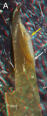
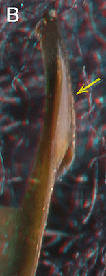
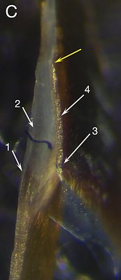
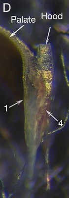
Figure. S. spirula, male, 31 mm ML. T0P: Side view showing where the longitudinal cut was made. BOTTOM: A - Oral-medial view of the longitudinal cut. B - A more oral view showing the Palatoshoulder Ridge (lighter color) overlapping the Hyaline Matrix of the shoulder. C - Photomicrograph (2D), oral view of the longitudinal cut taken perpendicular to the plane of the cut. D - Same beak; photomicrograph (2D) of the cross-sectional cut, posterior view taken perpendicular to the plane of the cut.
ARROWS: 1 - Palatoshoulder Ridge posteriorly at the surface of the cut. 2 - Palatoshoulder Ridge seen through the hyaline matrix as it doesn't quite reach the level of the cut at this point. 3 - Follow the direction of the arrow onto the Hyaline matrix to see the posterior edge of the cut surface. 4 - Shoulder Blade. Yellow arrows - Point to same spot on all beaks but D.S. spirula has a distinct, but reduced Palatoshoulder Ridge with Hyaline Matrix forming the middle part of the Shoulder. The Shoulder Blade appears, from both cuts, to be of simple (single component) construction. Fig. C, 3 shows the heavy pigment dorsal and posterior to the Hyaline Matrix is beneath the plane of the cut. Nevertheless, the means by which the posterior wall of the Hyaline Matrix becomes pigmented is uncertain. The Shoulder Blade lacks any external covering. Note that the Hyaline Matrix is in early stages of pigmentation which makes interpretation of structure more difficult.
ORDER UNCERTAIN
Superfamily Bathyteuthoidea
BATHYTEUTHIDAE: one genus
UPPER BEAK CHARACTERISTICS
- Step: Absent.
- Shoulder Blade: Thin; outer-most major layer of Shoulder; not partially covered laterally by thin layer of hyaline material.
- Shoulder Blade: Consists of one component.
- Hyaline Matrix: Forms Middle Part of Shoulder.
- Palatoshoulder Ridge: Present but reduced; extends onto the Palate.
- Pigmentation of posterior surface of Hyaline Matrix: Unknown.
- Yellow line: Absent.
Phylogenetic relationships. On the basis of morphology Bathyteuthidae and Chtenopteryx have been placed together in a superfamily. These families contain characteristics of both the Sepioidea and Oegopsida. Molecular studies (Lindgren, 2010 and Lindgren, et al., 2012) have confirmed the sister relationship of these families and have established the position of the Bathyteuthoidea as a sister taxon to the Oegopsida. Their closest relative within the Sepioidea/Spirulida/Idiosepiidae complex, however, is unresolved (Lindgren, 2010 and Lindgren, et al., 2012).
Bathyteuthidae: Structure
- EXTERNAL FEATURES
xxxxxxxxx
Bathyteuthis sp., 33 mm ML, 0.9 mm URL. LEFT - Oblique view.
Bathyteuthis berryi, 53 mm ML, 0.8 mm URL, Hawaiian waters. MIDDLE - Side view. RIGHT - Oblique view.
The mature (?) Bathyteuthis berryi beak (Middle, Right) shows no step in the Shoulder. It has what appears to be a Palatoshoulder Ridge (arrow) but it looks much like the shoulder of various mature sepiolids that retain little of the structure of the Shoulder. This, however, is not the case (see E, F below). The Shoulder Blade seems to have a simple construction. No hyaline material is present lateral to the Shoulder Blade or medial to the Palatoshoulder Ridge. The shoulder of the immature (?) Bathyteuthis sp.(Left) has a narrow but distinct Palatoshoulder Ridge (arrow).
- INTERNAL STRUCTURE: Longitudinal and cross-sectional cuts through the Shoulder. Click on an image to view larger version & data in a new window
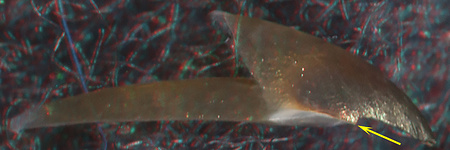
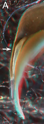
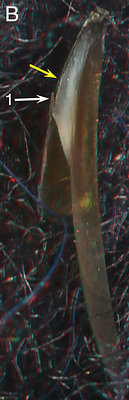
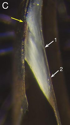
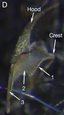
Figure. Bathyteuthis sp., sex ?, presumably immature, 33 mm ML, 0.9 mm URL. TOP: Left - Side view showing where the longitudinal cut was made. Right - Posterior view of the cross-sectional cut (3D); same view as the 2D photo in Fig. D. BOTTOM: A - Posterior view of left side. B - Oral-medial view. C - Photomicrograph (2D) of the longitudinal cut, oral view taken perpendicular to the plane of the cut. D - Photomicrograph (2D) of the cross-sectional cut taken perpendicular to the plane of the cut.
ARROWS: A - Arrow shows approximate level of longitulinal cut. B, 1 - Broken piece of shoulder blade due to poor cut. Note that this piece is missing in Fig. C. C, 1 - The anterior end of the Palatoshoulder Ridge which is also the anterior end of the cut; much of the Ridge is buried within the Hyaline Matrix. C, 2 - Anterior end of the Ridge that lies within the plane of the cut. D, 1 - Palatoshoulder Ridge. D, 2 - Hyaline Matrix. D, 3 - Shoulder Blade. Yellow arrows - Point to same spot on all beaks where present
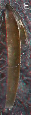
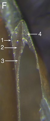
Figure. Bathyteuthis berryi, ? sex, presumably mature judging from the squids size and heavy beak pigmentation, 53 mm ML, 0.8 mm URL, with longitudinal cut through the shoulder. E - Oral-lateral view. F - Photomicrograph (2D) of the cut shoulder, oral-lateral view taken perpendicular to the plane of the cut.
ARROWS: 1 - Palatoshoulder Ridge. 2 - Hyaline Matrix with the medial face of the Shoulder Blade visible through the translucent Matrix. 3 - Pigmented posterior covering of the Hyaline Matrix. 4 - Shoulder Blade.The cuts of both beaks were taken close to the Jaw Angle and, therefore, show the Palatoshoulder Ridge which is narrow (dorsal-ventral axis) at least in the younger beak (Left image, table of photos). The Shoulder Blade is simple (i.e., single component) in the younger beak but thicker, and possibly more complex in the older beak. The added thickness, presumably, is associated with the posterior pigment cover of the Hyaline Matrix.


