*** Abraliopsis falco: Integumental photophores of the head and anterior mantle ***


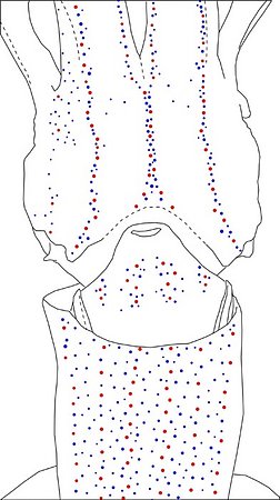
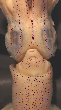
Figure. Ventral views of the integumental photophores of Abraliopsis falco, mature male, 37 mm ML, off Baja California. Left - Photograph of the preserved squid. Middle - Outline drawing with all integumental photophores represented by colored dots. Right - Photograph withcolored dots superimposed on integumental photophores. Red dots - Complex photophores. Blue dots - Non-complex photophores. Images by R. Young.
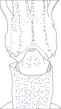
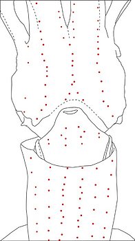
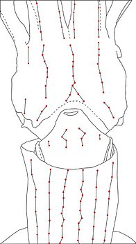
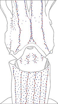
Figure. Ventral views of the same squid. Top left - Drawing showing only the non-complex (blue) photophores. Top middle - Drawing showing only the complex (red) photophores . Top right - Same drawing with lines connecting red photophores to aid comparisons of photophore patterns between species. Bottom - Drawing showing all photophores and lines showing red photophore patterns. Drawings by R. Young.
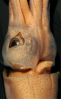
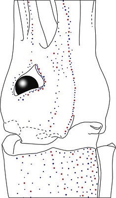
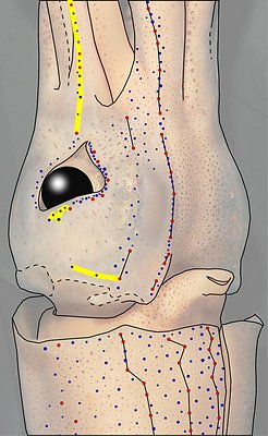
Figure. Ventral views of the integumental photophores of Abraliopsis falco, mature female, 41 mm ML, off Baja California. Left - Photograph of the preserved squid. Middle - Outline drawing from photograph with all integumental photophores represented by colored dots. Right - Outline drawing over dimmed photograph with all integumental photophores represented by colored dots, and with lines connecting some photophores. Red dots - Complex photophores. Blue dots - Non-complex photophores. Lines connecting photophores assist in understanding the organization of the photophores. Black lines - Patterns based on red photophores. Yellow line - Pattern based on red and blue photophores. Images by R. Young.
*** Abraliopsis falco: White photophores of the funnel groove ***
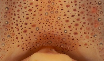
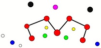
Figure. Ventral view of the funnel-groove region of the head of A. falco, 41 mm ML, mature female. Top - Photograph of the preserved squid showing photophores of the funnel groove and surrounding tissue. Middle - In the photograph, white dots are placed over the "white" photophores of the funnel groove. Black dots are placed over the two most posteromedial "red" photophores of the ventral head to mark the anterolateral edges of the funnel groove. Bottom - Same pattern of dots as in photographs but dots are colored. Colored dots, lines - Aid in comparison of patterns between species. Open dots - Represent photophores that are either of uncertain status as "white" photophores or have variable presence. Images by R. Young. Comparison of white photophore patterns for all Abraliopsis species can be found here.
Comments: Summary of photophore distributions. (photophore definitions found here)
ARMS:
Medial Arm IV Series - Continuous; reaches about half of arm length.
Central Arm IV Sector - Photophores absent.
Lateral Arm IV Series - Discontinuous; reaches arm tip.
Lateral Membrane Series of arms IV - Reaches about one third of the arm length.
Arm III Series - Discontinuous; extends nearly to arm tip.
HEAD - Typical three photophore series on central-ventral head with:
Median Head Series - At least 5 "red" photophores in posterior caret. Anteriorly red photophores in a somewhat irregular, single series.
Median Head Sector - Absent.
Medial Head Sector - A few (ca.3-4) scattered blue photophores occasionally present at anterior end of sector in large squid, otherwise photophores absent.
Lateral Head Series - Single, nearly straight, series of red photophores.
Window Sector - Scattered, blue photophores anterolaterally, otherwise photophores absent.
First Lateral Window Series - Two segments broadly separated by mostly-single series of blue photophores: Each segment with 2 red photophores. Apparently no red photophores lie on lateral membrane series of arm IV - needs confirmation.
Second Lateral Window Series - Absent.
Eyelid Series - Red photophores only on ventral half of eyelid. Posterior Eyelid Series with two large blue photophores displaced slightly posteriorly from eyelid series; more ventral of two, larger and more posterior. Formula: RbbBbB.
Occipital Series - Rbb.
Lateral Funnel-groove Series - 3 (1+2) or 4 (2+2) red photophores, in two segments well aligned.
White Patch (funnel groove) - Complex-2 Pattern (red, green, blue; + yellow, maroon at larger ML).
FUNNEL:
Medial Patch - "Nose" pattern of 4 "red" photophores.
Lateral Patch - Two red photophores.
MANTLE - Typical six photophore series on ventral mantle with:
Median Sector - Broad, but with broadly scattered blue photophores often obscuring central bare strip both anteriorly and posteriorly.
Medial Mantle Series - Slightly irregular alignment in anterior half of mantle.
1st Lateral Mantle Sector - Blue photophores scattered throughout sector.
2nd Lateral Mantle Sector - Fewer blue photophores scatterred throughout sector.
Lateral and Mantle-angle Series - Both very distinct with virtually straight alignment (alignment anteriorly veers laterally along as mantle broadens).
Comments
The above information has been extracted from photographs and from Young (1972).


