I. External structure.

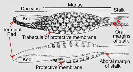
Figure. Tentacular club of Loligo forbesi. Top - Oral view. Bottom - Aboral view. Drawings from Naef 1921-23.
Features:
- Carpus: Absent.
- Club divisions: Manus, dactylus and terminal pad.
- Club shape: Expanded manus with narrow dactylus generally having a dorsal curvature.
- Sucker series: Four, regular, longitudinal series except for few suckers at proximal end of club. Four suckers comprising a transverse row are arranged at oblique and acute angles to the axis of the club rather than at a right angle.
- Trabeculate, protective membranes: One trabecula per lateral sucker; dorsal membrane low to virtually absent on distal half of dactylus; dorsal and ventral membranes nearly join at proximal end of club. Trabeculae of ventral membrane may vary in shape from those of dorsal membrane.
- Oral margins of stalk: Present; may be weak or prominent; without trabeculae; located near oral midline of stalk; continuous with protective membranes of club; extend length of stalk.
- Aboral margin of stalk: Present; may be prominent; positioned slightly dorsal of mid-aboral line; without significant musculature; not continuous with keel (ends distally about where keel begins); proximally extends full length of stalk.
- Keel: Extends almost full length of club. At proximal end muscular keel aligned with non-muscular aboral margin in approximate mid-aboral position; distally keel gradually shifts to dorsal position along dactylus.
- Terminal pad: Present; sometimes difficult to distinguish from dactylus.
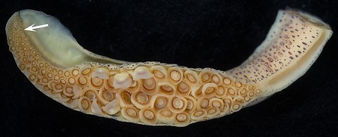
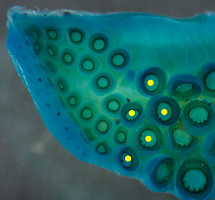
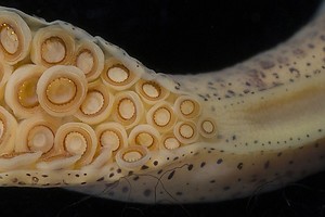
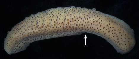
Figure. Sepioteuthis sp., 64 mm ML, Palau. Top - Oral view of club. Arrow marks separation of dactylus and terminal pad. Middle-left - Magnification of the terminal pad. The tip of the club is curved toward the camera and lightly stained with methylene blue stain. The yellow dots mark the terminal suckers of the dactylus which are toothed in contrast to the smooth-ringed suckers of the terminal pad. Note the very distinctive terminal pad on this species. Middle-right - Magnification of the proximal end of the club showing protective membranes continuing onto the stalk as oral margins. Bottom - Ventroaboral view of club. Arrow indicates proximal end of the keel and distal end of the aboral margin of the stalk. Photographs by R. Young.
II. Internal structure: Skin, keel and suckers removed.
- Dorsal view: The lateral sucker-stalks are greatly enlarged on the mid-manus even though their suckers are similar in size to those of the medial suckers. The size of the lateral sucker-stalks decreases abruptly at the dactylus as does the trabeculate protective membrane.
- Ventral and slightly aboral view: This opposite view shows the enlarged suckers of the mid-manus decreasing in a uniform manner to the tip of the dactylus along with the trabeculate protective membrane.
- The keel has been mostly removed leaving only the attachment region; nevertheless, the position of the keel can be seen changing from dorsal along the dactylus to aboral at the proximal region of the manus.
- Oral view: The sucker stalks of medial suckers lie in deep pockets that are joined by the "open canal" (see next section). Musculature is complex; trabeculae are more strongly attached to the more distal sucker stalks (this is most apparent among the dorsal trabeculae in the photograph below).
Click on an image to view larger version & data in a new window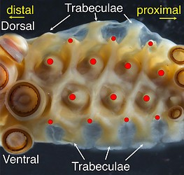
Figure. Sepioteuthis sp., oral view of the manus with some suckers removed showing the complex arrangement of surface muscles. Note the deep pockets surrounding the medial suckers.
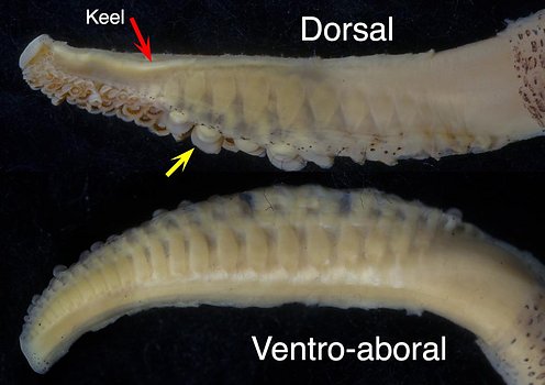
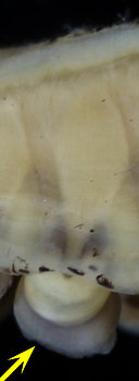
Figure. Sepioteuthis sp., 64 mm ML. Left - Dorsal and ventro-aboral views of the club with the skin removed to show sucker stalks and the keel position; most of the keel (red arrow) has been cut-off. Note that the sucker stalks and protective membrane virtually disappear along the dorsal margin of the dactylus compared to the ventral side of the dactylus. Right - Higher magnification (compare left yellow arrows in left and right images) showing relationship between sucker and sucker stalk. Photographs by R. Young.
III. Internal structure: Cross-section of the mid-manus; stalk.
- Organization: Manus somewhat bilaterally symmetrical with long stalks to lateral suckers and short stalks to medial suckers (irrespective of sucker sizes). (See "Basal clubs: Introduction" for comments on use of "bilateral symmetry.") Oral surface of manus basically flat when in relaxed state.
- Trabeculate protective membranes: Dorsal and ventral membranes large and equally developed.
- Core shape: Circular.
- Keel: Attached slightly dorsal to mid-aboral side of club core.
- Attachment of sucker stalks and trabeculae: Sucker stalks attach mostly to club core with some attachment to other sucker stalks; trabeculae attach to both the sucker stalks and club core. The complex musculature of the club is poorly understood.
- Open canal: Present; zig-sags along the midline beneath the oral surface of the club and opens onto oral surface at the deep pockets surrounding the medial sucker-stalks.
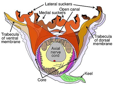
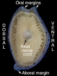
Figure. Sepioteuthis sp., 31 mm ML, Palau. Left - Cross-section through the manus showing the basic structure of the club core, and the attachment of the keel, sucker stalks and trabeculae of the protective membranes. Most of the core outside the axial nerve cord is composed of muscles. Dots on drawing areas indicate primarily longitudinal muscles. Other colors and fill-lines indicate more complex musculature. The red dot on the aboral side of the nerve cord is the major artery of the club; the blue dot oral of the nerve cord is the major vein of the club. The drawing, by R. Young, is a compilation taken from histological cross-sections. Right - Cut using a scapel through the tentacular stalk just proximal to the club.
IV. Internal structure: Cross-section of the mid-dactylus.
- Organization: Strong asymmetry with very long stalks for the ventrolateral suckers grading to small stalks for dorsolateral suckers. Oral surface of dactylus basically flat, when in relaxed state, and in same plane with that of manus.
- Trabeculate protective membranes: Ventral trabecula and membrane well developed; dorsal counterparts greatly reduced.
- Core shape: Circular; somewhat tilted ventrally compared to manus.
- Keel: Large; attached to dorsal side of core; oral surface (when relaxed) parallel to that of combined suckers.
- Attachment of sucker stalks and trabeculae: Except for the asymmetry, similar to that of mantle.
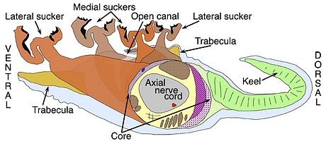
Figure. Sepioteuthis sp., cross-section through the dactylus showing the basic structure of the club core, and the attachment of the keel, sucker stalks and trabeculae of the protective membrane. The drawing, by R. Young, is a compilation taken from histological cross-sections.
General comments:
Note the strong contrast in the shape of the cross-sections between the manus and dactylus.


