- Arms
- Arms I 50-55% of ML
- Arms III 85-90% of ML
- Arms IV 150-170% 0f ML
- Large arm suckers with 22-26 pointed, triangular teeth, in contact at bases, over distal 2/3 of ring.
- Largest suckers not globular.
- Tentacular clubs
- Club length 28-35% of ML
- Suckers with 7-8 pointed teeth, in contact at bases, over distal 1/2 of ring; no enlarged central tooth.
- Suckers with stalks in two distinct parts; stalks of lateral suckers about equal in length to stalks of medial suckers.
- Protective membranes
- Membranes in three distinctive sets
- Proximal set with 7-12 trabeculae (14-18% of club length).
- Middle set without distinct trabeculae but opposite 9-12 marginal suckers (32-36% of CL).
- Distal set with 20-21 trabeculae (46-54% of CL).
 Click on an image to view larger version & data in a new window
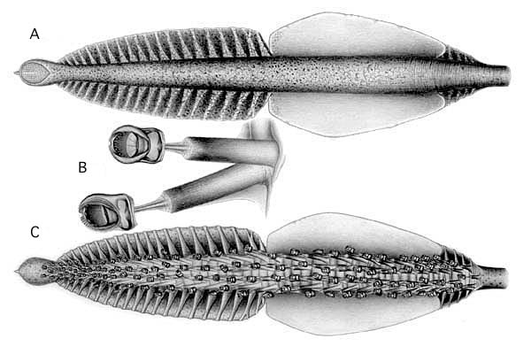
Figure. Tentacular club of C. joubini. Left - A - Aboral view of club. B - Club suckers and stalks. Note that stalks are approximately equal in size, the pleates are absent, and each stalk is in two parts (thick basal segment and thin distal segment). C - Oral view of club. Note short proximal section defined by the protective membranes. Drawings by A. D. Hart.
Click on an image to view larger version & data in a new windowClick on an image to view larger version & data in a new window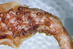
Figure. Oral view of the proximal end of the tentacular club of C. joubini showing the short, proximal set of protective membranes. Photograph by R. Young.
- Head
- Head length 34-37% of ML.
- Head length 34-37% of ML.
- Funnel
- Tragus and antitragus of the funnel-locking apparatus large and overhang beyond their bases; tragus high and narrow.
- Photophores
- Eyeball with oval organs in 2 series: lateral series= 5 or 6 organs; 1(enlarged)+4(or 3)+1; medial series = 5 or 4+1. Formula reads from posterior to anterior.
- Viscera: two, large photophores.
- Club-tip photophore of moderate size with small papilla.
- Distal-most cushion-like photophore partially embedded in aboral surface of stalk proximal to club.
- Pigmentation*
- Club-tip photophore with chromatophores only.
- Most club pigmentation in chromatophore organs; epithelial pigment and scattered chromatophores on aboral surface of proximal portion of club.
- Club sucker stalks with epithelial pigment but not pleated; pigmentation diminishes then disappears from stalks in distal portions of club.
- Buccal membrane unpigmented.
- Olfactory organ with a few chromatophores on sensory bulb.
Scanning electron micrographs of the arm and club suckers can be seen here.
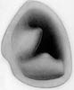
Figure. Frontal view of the funnel locking-apparatus of C. joubini. Drawing by A. D. Hart.
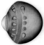
Figure. Left -Ocular photophores of C. joubini. Right - Visceral photophores of C. joubini.
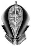
Figure. Club-tip photophore of C. joubini with the lids cut and spread open. Note the longitudinal stripe down the center of the photophore. Drawing by A. D. Hart.
Comments
The holotype was described by Joubin (1933) as having a single abdominal photophore. Our re-examination of the type shows that two photophores are actually present. Also, contrary to Joubin's description, the arrangement of photophores on the eye is 1+4+1, 4+1. The above characters are based mostly on two specimens: the 99 mm ML holotype and a 107 mm ML squid from off Bermuda (sex unknown). The intermediate protective membrane on the holotype has cleared slightly due to preservation. The membrane consists of a solid mass of undivided trabeculae with a membrane visible only near the rounded tips of the trabeculae.
*The density and type of pigmentation varies with the size/age of the squid with smaller squid having less pigmentation and more often in chromatophores.


