Back to Profile Page

Tree of Life Media Contributed By Brian S. Leander

<< < 1 2 3 4 5 6 7 > >>
ID Thumbnail Media Data
30381
Comments Illustration showing the organization and main components of the euglenid pellicle.
Reference Modified from Leander, B. S., H. J. Esson, and S. A. Breglia. 2007. Macroevolution of complex cytoskeletal systems in euglenids. BioEssays 29:987-1000.
Creator Heather Esson and Brian Leander
Copyright © 2007 Heather Esson and Brian Leander
Image Use restricted
Attached to Group Euglenida: view page image collection
Title strip.jpg
Image Type Diagram
Image Content Ultrastructure
Subject Histology, Morphology
ID 30381
30387
Specimen Condition Dead Specimen
Copyright © Brian S. Leander
Image Use restricted
Attached to Group Euglenida: view page image collection
Title sems2.jpg
Image Type Photograph
Image Content Specimen(s), Ultrastructure
Technical Information Scanning electron micrographs
ID 30387
30388
Comments Scanning electron micrographs showing the relative number of pellicle strips and the size differences between a small bacterivore (left, with 4 pellicle strips) and a large euglenophyte (right, with 48 pellicle strips).
Specimen Condition Dead Specimen
Copyright © Brian S. Leander
Image Use ToL use only
Attached to Group Euglenida: view page image collection
Petalomonas: view page image collection
Lepocinclis: view page image collection
Title size.jpg
Image Type Photograph
Image Content Specimen(s), Ultrastructure
Technical Information Scanning electron micrograph
ID 30388
30441
Scientific Name Distigma, Eutreptia, Euglena
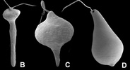
Comments metaboly in different euglenids: (B) Distigma, (C) Eutreptia and (D) Euglena
Specimen Condition Dead Specimen
Copyright © 2004 Brian S. Leander
Image Use ToL use only
Attached to Group Euglena: view page image collection
Distigma: view page image collection
Eutreptia: view page image collection
Title metaboly.jpg
Image Type Photograph
Image Content Specimen(s), Ultrastructure
Technical Information scanning electron micrographs
ID 30441
30442
Scientific Name Monomorphina (A), Phacus (B-C), Lepocinclis (D-E), Rhabdomonas (F)
Comments Scanning electron micrographs of photoautotrophic and primary osmotrophic euglenids that have independently lost the ability to undergo metaboly
Specimen Condition Dead Specimen
Copyright © Brian S. Leander
Image Use restricted
Attached to Group Monomorphina: view page image collection
Lepocinclis: view page image collection
Phacus: view page image collection
Rhabdomonas: view page image collection
Title rigid.jpg
Image Type Photograph
Image Content Specimen(s), Ultrastructure
Technical Information Scanning electron micrographs
ID 30442
30446
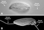
Comments Images showing the heterodynamic orientation of the dorsal (anterior) and ventral (posterior) flagella in gliding (phagotrophic) cells.
Image Use creative commons This media file is licensed under the Creative Commons Attribution-NonCommercial License - Version 3.0.
Copyright © 2008 Brian S. Leander
Attached to Group Euglenida: view page image collection
Title flag3.jpg
Image Type Photograph
Image Content Specimen(s), Ultrastructure
Technical Information Light micrograph (A) and scanning electron micrograph (B)
ID 30446
30816
Scientific Name Stephanopogon minuta
Creator Image taken by Naoji Yubuki
Image Use creative commons This media file is licensed under the Creative Commons Attribution-NonCommercial License - Version 3.0.
Copyright © Brian S. Leander
Attached to Group Stephanopogon: view page image collection
Title stephanopogon1.jpg
Image Type Photograph
Image Content Specimen(s)
ID 30816
30867
Comments eukaryovorous cell showing the feeding rods
Copyright © Brian S. Leander
Image Use ToL use only
Attached to Group Euglenida: view page image collection
Title feed1.jpg
Image Type Photograph
Image Content Specimen(s), Ultrastructure
Technical Information light micrograph
ID 30867
32139
Comments Organization of the euglenid feeding apparatus: relationship of the feeding rods with the flagellar pocket (the anterior tip of the cell has been cut to show a transverse view of the apparatus).
Reference modified from Leander, B. S., H. J. Esson, and S. A. Breglia. 2007. Macroevolution of complex cytoskeletal systems in euglenids. BioEssays 29:987-1000.
Copyright © Brian S. Leander
Image Use ToL use only
Attached to Group Euglenida: view page image collection
Title feedingrods.jpg
Image Type Diagram
Image Content Specimen(s), Ultrastructure
ID 32139
30876
Comments Two cells that glide along substrates showing a straight anterior flagellum (A) and a thick recurrent or posterior flagellum that trails the cell (B)
Copyright © Brian S. Leander
Image Use ToL use only
Attached to Group Euglenida: view page image collection
Title general1.jpg
Image Type Photograph
Image Content Specimen(s), Ultrastructure
Technical Information Light micrograph
ID 30876
Please note: Most images and other media displayed on the Tree of Life web site are protected by copyright, and the ToL cannot act as an agent for their distribution. If you would like to use any of these materials for your own projects, you need to ask the copyright owner(s) for permission. For additional information, please refer to the ToL Copyright Policies.

Contribute to the ToL

ToL Contributions

Ways to Contribute

Use of Contributions

Scientific Content

Building Treehouses

Images/Media

  go to the Tree of Life home page
top