Several species have been placed in the Mastigoteuthidae that we consider species dubia or synonyms of known species. These species are briefly described here in the following order: Mastigoteuthis latipinna, Mastigoteuthis islini, Mastigoteuthis inermis, Mastigoteuthis okutanii and Chiroteuthoides hastula.
Mastigoteuthis latipinna (Sasaki, 1916)

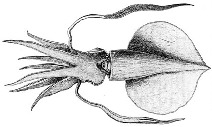
Figure. Ventral view of M. latipinna, holotype. Drawing from Sasaki (1916).
Introduction
M. latipinna (originally Sasaki created the mastigoteuthid genus Idioteuthis for this species) was described on the basis of a single large squid (238 mm ML), the largest of any species of mastigoteuthid known at the time. The eyes were described as being greatly different in size as seen in the illustration above. However, Sasaki, presumably believed that the apparent mismatched eyes resulted from damage as he did not site this difference when comparing his species with the similar M. cordiformis.
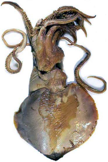
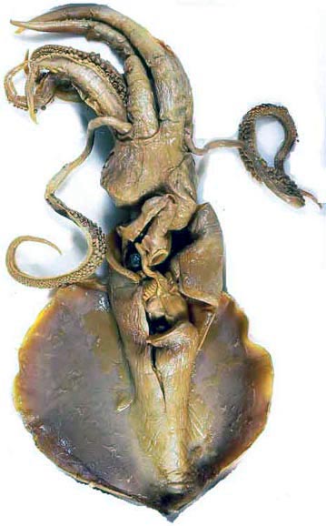
Figure. Dorsal and ventral views of M. latipinna, holotype. Photographs by A. Salcedo-Vargas.
Characteristics
- Arms
- Arm formula: 4>2>3>1. Arms IV about 2/3 of ML.
- Arm suckers number: arms I - III 70 pairs; arms IV - 65 pairs.
- Horny ring smooth, sometimes with horny lumps. [The latter is a preservation artifact]
- Tentacles
- Club distal half of tentacle; a little flattened proximally, cyclindrical distally; broad, trabeculate protective membranes border both sides of club.
- Sucker numbers in a transverse row on club increase from 4 at base to 24 distally.
- Proximal club suckers about same size as largest arm suckers; suckers become minute distally.
- Horny ring of proximal club sucker with about 10 blunt, minute teeth on distal margin; distal club suckers with about 25 conical but blunt teeth on distal four-fifths of margin with those more distal larger. Click on an image to view larger version & data in a new window
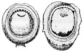
Figure. Oral view of tentacular club suckers of M. latipinna, holotype. Left - Distal sucker. Right - Proximal sucker. Drawings from Sasaki (1916).
- Head
- "Nuchal cartilage panduriform, narrowed in the middle, and traversed by a longitudinal median ridge with a groove along the crest."
Click on an image to view larger version & data in a new window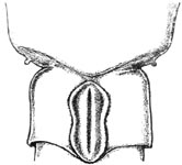
Figure. Frontal view of the nuchal cartilage of M. latipinna, holotype. Drawing from Sasaki (1929).
- Funnel
- Funnel locking apparatus: "... tragus and antitragus both faint and the latter being by far the fainter." Click on an image to view larger version & data in a new window
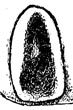

Figure. Frontal and medial-frontal views of the funnel locking-apparatus of M. latipinna, holotype. Drawing from Sasaki (1929); photograph by A. Salcedo-Vargas.
- Funnel locking apparatus: "... tragus and antitragus both faint and the latter being by far the fainter."
- Mantle and skin
- "smooth skin."
- Fins
- "... about five-sixths as long as the mantle, ... combined outline nearly circular... .
- Photophores
- Photophores absent

Figure. Oral view of arm suckers of M. latipinna, holotype . Drawings from Sasaki (1916).
Figure. Oral/lateral view of the tentacular club of M. latipinna, holotype. Photograph by A. Salcedo-Vargas.
Comments
Above description and quotes are from Sasaki (1929). Sasaki (1929) describes a small (90 mm ML) M. cordiformis in the same paper with the large M. latipinna. The differences between these specimens are probably a result of the size differential and damage. Slight differences in sucker dentition exist between forms of M. cordiformis from different regions (see the ToL page for this species). Whether or not these will eventually distinguish separate species is unknown. If they do, M. latipinna is the available name for the Japanese form. Until this is demonstrated, we consider M. latipinna to be a junior synomyn of M. cordiformis.
Distribution
Type locality: Sagami Sea, outside the Okinose bank, Japan.
References
Sasaki, M. 1916. Notes on Oegopsid Cephalopods Found in Japan. Annotationes Zoologicae Japonenses, 9(2):89-120.
Sasaki, M. 1929. A Monograph of the Dibranchiate Cephalopods of the Japanese and Adjacent Waters. Journal of the College of Agriculture, Hokkaido Imperial University, 20(supplement):357 pages.
Mastigoteuthis islini MacDonald and Clench, 1934
Introduction
M. islini was described on the basis of a single, badly damaged squid that lost both tentacles and apparently its skin during capture off the NE coast of the U.S.A.Characteristics
- Arms
- Arm suckers. "The spines of the chitinous rim are 13 in number, the median spine most strongly developed. Ventraly along the rim these sharp pointed spines are followed on each side by six blunt spines, the median ventral portion of the rim being slightly crenulated. The suckers have a short stalk but the stalks are located on tall, truncated, cone-shaped, gelatinous swellings..." Click on an image to view larger version & data in a new window
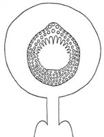
Figure. Oral view of sucker from arm IV of M. islini. Drawing from MacDonald and Clench (1934).
- Tentacles
- Tentacles lost during capture.
- Funnel
- Funnel locking-apparatus with "...the tragus well developed, but no trace is found of the antitragus." Click on an image to view larger version & data in a new window
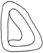
Figure. Frontal view of the funnel locking-apparatus of M. islini. Drawing from MacDonald and Clench (1934).
- Funnel locking-apparatus with "...the tragus well developed, but no trace is found of the antitragus."
- Mantle and skin
- Tubercules presumably absent.
- Fins
- "The fin is definitely elliptical in shape and relatively very large, being about three-quarters of the length of the mantle." Click on an image to view larger version & data in a new window
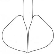
Figure. Ventral view of fins of M. islini. Drawing from MacDonald and Clench (1934) labeled "dorsal view of fin." The anterior mantle margin is not shown but exists at the end of the mantle lines according to the measurements.
- "The fin is definitely elliptical in shape and relatively very large, being about three-quarters of the length of the mantle."
- Photophores
- "We have been unable to find luminescent organs on th eyes or on any other part of the external surface of this species."
- Measurements
- Length to base of arms IV - 59 mm; width of eyes - 10 mm; mantle width - 15 mm; mantle length - 52 mm; length of fin - 35 mm; width of fin - 50 mm; length arm I - 36 mm; length arm II - 40 mm; length arm III - 42 mm; Length arm IV - 70 mm.
Comments
Quotations above are from MacDonald and Clench (1934). The holotype is located in Mus. Comp. Zool., No. 98,967. Nesis (1987) suggested that this species might be a synomyn of Mastigoteuthis atlantica presumably due to the absence of integumental photophores. We suspect that the absence of photophores is due to damage and consider M. islini to be a "species dubia."
Distribution
Type locality - 39° 04'N, 71° 29'W, North Atlantic off the NE coast of the United States.
References
MacDonald, R., and W.J. Clench. 1934. Descriptions of a new genus and two new species of squids from the North Atlantic. Occasional Papers of the Boston Society of Natural History, 8:145-152.
Mastigoteuthis inermis Rancurel, 1972
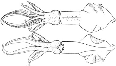
Figure. Dorsal and dventral views of M. inermis, holotype. Drawing from Rancurel (1972)
Introduction
Rancurel (1972) based the description of M. inermis on a single specimen, 142 mm ML, with intact tentacles.
Diagnosis
A Mastigoteuthis ...
- with flask-shaped funnel locking-groove.
- without photophores.
- with fin length greater than fin width.
Characteristics
- Arms
- Basal arm suckers largest (Rancurel, 1972).
- Arms (including arms IV) average 72 pairs of suckers.
- Arm suckers with smooth inner rings (Rancurel, 1972). Click on an image to view larger version & data in a new window
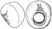
Figure. Lateral and oral views of arm suckers of M. inermis. Drawings from Rancurel (1972).
- Tentacles
- Club length 58% of tentacle length (Rancurel, 1972).
- Club suckers 0.06 mm in diameter (Rancurel, 1972).
- Club suckers with smooth inner rings (Rancurel, 1972). Click on an image to view larger version & data in a new window
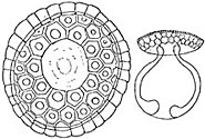
Figure. Oral and side views of club suckers of M. inermis. Drawings from Rancurel (1972).
- Head
- Nuchal cartilage from Rancurel, 1972 Click on an image to view larger version & data in a new window
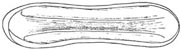
Figure. Dorsal view of nuchal cartilage of M. inermis. Drawings from Rancurel (1972).
- Nuchal cartilage from Rancurel, 1972
- Funnel locking-apparatus
- Groove of funnel component of locking-apparatus with flask-shape (Rancurel, 1972). Click on an image to view larger version & data in a new window
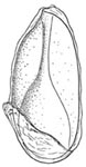
Figure. Ventral view of funnel locking-apparatus of M. inermis. Drawing from Rancurel (1972).
- Groove of funnel component of locking-apparatus with flask-shape (Rancurel, 1972).
- Mantle
- Tubercules absent from mantle and elsewhere.
- Fins
- Fin length (including 18 mm tail) 67% of ML; fin width 53% of ML (Rancurel, 1972).
- Photophores
- Integumental and eyelid photophores absent (Rancurel, 1972).
- Pigmentation
- Color brown-red (Rancurel, 1972).
- Measurements
- Measurements and counts according to Rancurel (1972) are: Sex - Female; Mantle length - 142 mm; Mantle width - 32 mm; Fin length - 95 mm; Fin width - 75 mm; Arm I, length - 63 mm; Arm II, length - 75 mm; Arm III, length - 67 mm; Arm IV, length - 128 mm; Arm I-III, ave. sucker no. - 150; Arm IV, sucker no. - 150; Tentacle length - 240 mm; Club length - 140 mm.
Comments
This description is taken from Rancurel (1972). M. inermis differs from M. magna primarily in the greater length of the fin relative to its width. However, there is a large variation in fin measurements in mastigoteuthids depending on the damage and state of contraction. We consider M inermis to be a junior synonym of the broadly distributed M. magna.
Distribution
Type locality: Eastern tropical North Atlantic, 25 miles south of Abidjan, Ivory Coast, Africa (ca 4.5°N, 4°W).
References
Rancurel, P. 1972. Mastigoteuthis inermis espèce nouvelle de Chiroteuthidae du Golfe de Guinée (Cephalopoda - Oegopsida). Bulletin de la Societe Zoologique de France, 97(1):25-34.
Mastigoteuthis okutanii (Salcedo-Vargas, 1997)
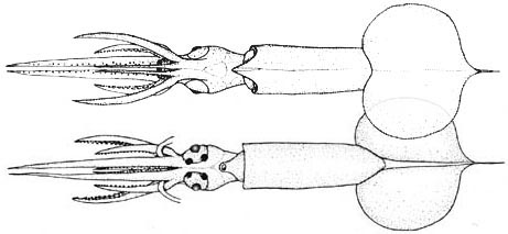
Figure. Dorsal and ventral views of M. okutanii, from the type series . Drawing from Salcedo-Vargas (1997).
Introduction
M. okutanii (originally described as Idioteuthis okutlanii) is based on four paralarvae that range in size from 15-18 mm ML which were missing their tentacles.
Diagnosis
A Mastigoteuthis ...
- with two photophores on each eyeball.
- with a western Indian Ocean habitat.
Characteristics
- Arms
- Arm suckers not described.
- Tentacles
- Missing.
- Funnel
- Funnel locking-apparatus large.
- Tragus and antitragus not present. Click on an image to view larger version & data in a new window

Figure. Frontal view of the funnel locking-apparatus of M okutanii. Drawing from Salcedo-Vargas (1997).
- Mantle
- Wart-like skin tubercules present but seen only around eyes.
- Photophores
- Two large, round, white photophores on ventral side of each eyeball.
- Integumental and eyelid photophores absent.
- Pigmentation
- Small chromatophores cover body and arms, mainly on ventral side.
- Measurements
Source Salcedo-Vargas, 1997 Salcedo-Vargas, 1997 Salcedo-Vargas, 1997 Salcedo-Vargas, 1997 Sex ? ? ? ? Mantle length 18 18 18 15 Mantle width 4 4 4 4 Fin length 10 9.5 9.5 10 Fin width 12 11 10 10 Head length 5.5 5.5 5.5 4.5 Head width 4 4 4 3.5 Arm I, length 4 4 4 3 Arm II, length 6 6 5.5 4 Arm III, length 4 4 4 3 Arm IV, length 9 9 -- 7.5 Tentacle length -- -- -- -- Club length -- -- -- -- Eye diam. / lens diam. 2.5 / 1.5 2.0 / 1.5 2.0 / 1.5 1.5 / 1.0
Comments
This description is taken from Salcedo-Vargas (1997). The only species previously known to have two ocular photophores is M. hjorti. Salcedo-Vargas (1997) observed, but didn't illustrate, a paralarva which had been identified as M. hjorti of the same size (ie, 16 mm ML) as M. okutanii in the South African Museum. This paralarva differed from the latter in eye size and fin width which indicated to Salcedo-Vargas that the two represented different species. Such differences could result from problems in determining accurate mantle lenghts. However, even if the two paralarvae represent different species, we do not know which paralarva belongs to M. hjorti. As a result, we have placed M. okutanii as a junior synonym of M. hjorti.
Distribution
Type locality: Indian Ocean off the coast of Somalia at ca. 51°N, 09°E, 0-100 m depth. This species is known only from the type locality.
References
Salcedo-Vargas, M. A. 1997. Cephalopods from the Netherlands Indian Ocean Programme (NIOP) - II. Mastigoteuthid lineage and related forms. Beaufortia, 47: 91-108.
Chiroteuthoides hastula Berry, 1920
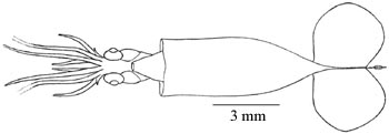
Figure. Ventral view of Chiroteuthoides hastula, holotype. Drawing from Berry (1920).
Introduction
Berry (1920) described a small collection of paralarvae from the north Atlantic. One of these was a single specimen of 10+ mm ML which he placed in the new genus and species Chiroteuthoides hastula. Berry was particularly struck by the very short arms III, a feature that we now know to be typical of Mastigoteuthid paralarvae.
Characteristics
- Arms
- Arms IV "...much the largest and longest, about two-thirds as long as the body (exclusive of fins and needle-like process..."
- Arms III "...excessively small, being between one-third ad one-half as long as the neighboring second pair."
- "Suckers minute, in two rows throughout."
- Tentacles
- Lost during capture.
- Funnel locking-apparatus
- "...their grooves simple."
- Mantle and skin
- Tubercules apparently absent.
- Fins
- "Fins nearly circular, about one-third as long as the sac-like part of the mantle.."
- Photophores
- Absent.
Comments
Quotations above are from Berry (1920). The single specimen is a 10+ mm ML (16.5 mm total length) paralarva. Young (1991) returned this species to the Mastigoteuthidae where it had earlier been placed by Naef (1923). Without chromatophore patterns, determining the subadult identy of this typical Mastigoteuthis paralarva will probably be impossible. We consider this to be a species dubia. The holotype is in the U.S. NMNH Cat. No. 338693.
Distribution
Type locality - 28°59'N, 69°22'W, western North Atlantic.
References
Berry, S.S. 1920. Preliminary diagnosis of new cephalopods from the western Atlantic. Proceedings of the United States National Museum, 58(2335):293-300, 1 plate.
Naef, A. 1923. Die Cephalopoden. Fauna e Flora de Golfo di Napoli, Monograph 35, 1(l) pt. 2:150-863.
Young, R. E. (1991). Chiroteuthid and related paralarvae from Hawaiian waters. Bull. Mar. Sci., 49: 162-185.


